Oral Precancerous Lesion Signals Smokers Must Watch For
In November 2023, I was waiting for a follow-up number in the oral mucosa department corridor of a tertiary hospital in Hangzhou. A man around 50 sat next to me; the smell of smoke seeped from the collar of his jacket. He pulled out his phone to show me a photo of the inside of his mouth — a gray-white patch on the left buccal mucosa with fuzzy edges and a few small reddish areas mixed in. He said he had noticed it for at least two months. At first he thought it was "smoke stains from smoking," gently brushed it with a toothbrush, applied Bingpeng Powder, and the white area seemed to fade a little, but came back thicker two weeks later.
After the doctor called his number, an examination was performed, and a biopsy was recommended that same afternoon. The pathology result came back: leukoplakia with epithelial dysplasia. Not cancer yet, but already in a range requiring serious follow-up and intervention. He later said something I still remember: "I stretched 'let me observe a few more days' into two months."
This article is written for smokers who are still in that "let me observe a few more days" phase. The focus is narrow: leukoplakia, erythroplakia, persistent ulcers, and when you should stop self-reassurance and go to the hospital.
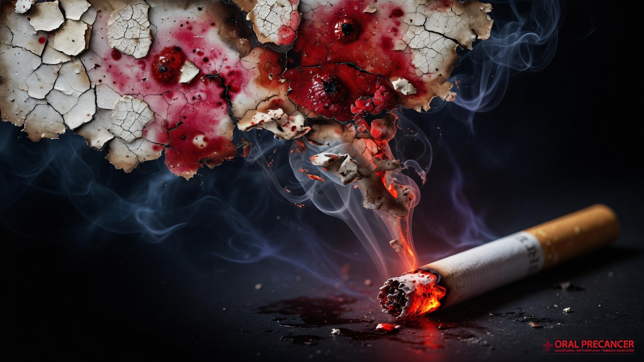
Let Me First State My Position
I do not advocate scaring every white spot in the mouth into cancer. Most mucosal leukoplakia does not immediately turn into oral cancer. Mayo Clinic and other institutions repeatedly state: many white patches themselves do not become cancerous, but leukoplakia elevates the risk of oral cancer, and cancer can also appear near white patches; mixed red-and-white spotty changes deserve more vigilance.
Nor do I advocate treating "white patches in smokers' mouths" as a normal working-life badge. Tobacco irritation is one of the most important risk factors for oral leukoplakia; long-term heavy alcohol consumption adds risk; smoking and drinking together is worse. Common expressions in domestic public science communication state: about 22% of oral cancers worldwide are directly related to smoking, and smokers' risk of oral cancer can be 5–10 times that of non-smokers. Specific multiples fluctuate by study population, but the direction is consistent — smoking is not background noise; it is one of the main causes.
My personal judgment has only three points:
- Persistent abnormality in color and texture is more important than "does it hurt." Many precancerous lesions are almost painless in early stages.
- Time matters more than folk remedies. Ordinary mouth ulcers heal within about 7–10 days; if it persists beyond about 2 weeks, treat it as "must be investigated" rather than trying another spray.
- Red spots and "reddening white patches" take priority over "simple whiteness." Quiet redness is often more dangerous than conspicuous whiteness.
Signal One: Oral Leukoplakia — Whiteness That Cannot Be Wiped Off Is Worth Noting
What It Looks Like
In professional terms, oral leukoplakia is roughly: a predominantly white plaque on the oral mucosa that cannot be scraped off, after excluding other diseases that can be clearly diagnosed (such as Candida infection, friction-induced keratosis, etc.). It may appear as:
- Gray-white or porcelain-white patches with clear or irregular borders;
- Flat surface, or resembling wrinkled paper, granular, or verrucous raised;
- When gently wiped with clean gauze or a cotton swab, cannot be wiped off (a "white membrane" that can be wiped off entirely should first consider fungal infection, but still needs a doctor's assessment, not self-diagnosis).
Common sites include the buccal mucosa, tongue dorsum, and gingiva; smoking-related irritation also often appears in areas repeatedly exposed to smoke. Leukoplakia on "high-risk anatomical zones" such as the tongue margin, ventral tongue, and floor of the mouth requires a higher level of follow-up.
How It Links to Smoking
Long-term exposure to irritants in tobacco promotes hyperkeratosis and abnormal epithelial cell proliferation. An early observation by Sun Zheng's team at Beijing Stomatological Hospital is frequently cited in science communication: among patients diagnosed with oral leukoplakia, the proportion of severe dysplasia and cancerous transformation in the smoking group was significantly higher than in the non-smoking group; the higher the pack-year smoking history, the steeper the risk (pack-year = packs per day × years). You don't need to memorize percentages — just remember the mechanism: dose and time rewrite the texture of the mucosa; "getting used to it" does not mean safety.
There is also a publicly reported data point: some studies state that those who smoke about 20 cigarettes per day have a considerable probability of developing "smoke stains"-type changes within several years; some lesions may continue to progress. Media reports have even stated that "the risk of cancerous transformation rises sharply after patches turn red." The numbers are not entirely consistent across different sources, but the operational takeaway is: watch stable white patches; if they turn red, break down, or harden, escalate immediately.
Homogeneous vs. Heterogeneous — I Fear the Latter More
- Relatively homogeneous leukoplakia: relatively uniform color, relatively flat surface. Still requires evaluation, but the overall risk spectrum is broader.
- Non-homogeneous / speckled leukoplakia: white mixed with red, granular surface, ulcers, marked thickening and hardening, worm-eaten edges. In publicly available sources, the malignant transformation rate of non-homogeneous leukoplakia can be significantly higher than homogeneous; when accompanied by moderate to severe epithelial dysplasia, "wait and see" is even less acceptable.
The range of overall malignant transformation rates for leukoplakia given in literature and science communication is very wide (0.13%–34%, also about 7%–15%, non-homogeneous even higher at 15%–40%, etc.), which means: individual risk must be assessed comprehensively based on site, type, pathology, and history of smoking, alcohol, and betel nut — you cannot comfort yourself with an average.
Typical Misjudgments I Have Seen
- Mistaking leukoplakia for "excessive internal heat" and taking three boxes of cooling granules.
- Using a hard-bristled toothbrush to "scrub off the whiteness," causing erosion, forming a pseudomembrane the next day, and thinking it was healing.
- On the third day of a smoking cessation app check-in, seeing the white patch still there and declaring "quitting is useless" — epithelial changes are measured in weeks and months, not hours.
In 2022, I helped a colleague with an 18-year smoking history do home comparison photography: the same side of the buccal mucosa, fixed phone distance about 15 cm, flash at a 45-degree angle, one photo every Sunday evening. By the fourth week he admitted "the edge looks rougher than the first photo." Visual memory is extremely unreliable — date-stamped photos are more reliable than gut feeling.
Signal Two: Oral Erythroplakia — Rare, But Turn the Volume to Maximum
What It Looks Like
Oral erythroplakia (erythroplasia) clinically often presents as: a bright red patch with relatively clear borders, the surface may be velvety smooth, sometimes slightly lower than the surrounding mucosa (thinner keratin layer, more translucent blood vessels). It can be entirely red, or red with tiny white speckles — the latter is frequently mentioned in medical literature as associated with more severe epithelial changes and must be judged by a specialist.
Common sites similarly "prefer" the ventral tongue, floor of mouth, soft palate, buccal mucosa, etc. It is rarer than leukoplakia, so it is more easily overlooked: patients looking in the mirror mainly check whether their teeth are yellow and rarely flip up their tongue to check the ventral surface.
Why I Give Erythroplakia Higher Priority Than Leukoplakia
The consensus direction from multiple professional sources is consistent: the malignant potential of erythroplakia is significantly higher than general leukoplakia. Some statements indicate a considerable proportion of erythroplakia cases already approach or reach severe dysplasia, carcinoma in situ, or even invasive carcinoma at initial diagnosis. Cleveland Clinic and other institutions also list smoking and tobacco chewing as important related factors for erythroplakia and emphasize it as a potentially precancerous condition that must be taken seriously.
My personal view is straightforward: if you only worry about "white spots," immediately add "red spots, red patches, and red-white mixed lesions" to your must-check list. Erythroplakia may not hurt — pain is not a safety valve.
How to Differentiate from Inflammation in Practice (For Self-Check Clues Only, Not a Substitute for Diagnosis)
- Redness caused by burns, aggressive brushing, or denture friction usually has a clear cause and should markedly improve within days after removing the cause.
- Red areas without identifiable cause, fixed in extent, persisting beyond 2 weeks, bleeding easily on touch, or with a relatively firm base should be given high-priority appointments.
After a health lecture in March 2024, an audience member privately mentioned a "persistent red patch like red ink" on their soft palate, which they had treated with iodine glycerin for ten days. I had them use their phone's rear camera on the spot, tilt their head back, and use a tongue depressor (or a clean stainless steel spoon handle) for photos. The photos showed clear borders, not diffuse congestion. They had a biopsy the second week. I cannot expand on the case details here, but the process illustrates one thing: the correct action for erythroplakia is evaluation and pathology, not household disinfectants.
Signal Three: Persistent Ulcers — Write the Two-Week Rule Inside Your Cigarette Pack
Ordinary Ulcers vs. Dangerous Ulcers
Recurrent aphthous ulcers are painful but usually self-limiting, most healing within about 1–2 weeks. The trouble with cancerous or precancerous ulcers is:
- No obvious healing trend after 2 weeks;
- Raised edges, depressed center, like a shallow crater;
- Firm or even "cartilage-hard" base on palpation — descriptions that frequently appear in clinical literature;
- Can be relatively painless, or the pain is disproportionate to the appearance;
- May be surrounded by leukoplakia or erythroplakia.
Oral health articles reprinted by Xinhua News Agency, China News Service, etc., in their "World No Tobacco Day" features, list "non-healing ulcers, mucosal color changes, lumps" as signals requiring vigilance and emphasize that ordinary ulcers heal in about 7–10 days, while those persisting beyond 2 weeks require medical attention. This is highly consistent with international oral cancer early screening messaging.
The Smoking-Specific Delay Script
I have summarized a high-frequency script that I encounter every year with version updates:
- Days 1–3: think it's internal heat or a bite injury;
- Days 4–7: spray watermelon frost, apply ulcer patches, switch to "shallow puffs, fewer puffs";
- Days 8–14: heal a bit then split open again, blame spicy food and staying up late;
- After day 15: use "I've had this before and it healed on its own" to suppress anxiety.
The problem: past self-healing experience does not guarantee this time. The mucosa can give different answers in different years.
Three Things You Must Stop Immediately
- Using cigarette butts to burn or alcohol swabs to harshly "disinfect" the area;
- Repeatedly picking at the white membrane or peeling scabs with fingers;
- Simultaneously smoking and drinking alcohol to irritate the wound while expecting it to heal according to textbook timelines.
Secondary but Not Ignorable Signals
These may not individually equal precancerous lesions, but when combined with the three above, the grounds for seeking medical attention become stronger:
| Signal | Self-Check Tip |
|---|---|
| -------- | --------------- |
| Unexplained lump or localized thickening in the mouth | Use finger pads to compare left and right symmetrical areas, record whether it is fixed and whether it is growing |
| Stiff tongue movement, newly appearing slurred speech | Compare with voice recordings from 1 month ago (many people have WeChat voice messages in their phones) |
| Unexplained bleeding or numbness | If it recurs after excluding brushing trauma and burns, seek medical opinion |
| Painless cervical lymph node enlargement | After bathing, touch both sides in front of the mirror for comparison; soybean-sized or larger, relatively firm texture requires attention |
| Dentures suddenly don't fit, loose teeth without periodontal explanation | Prompt dental evaluation, do not just adjust the lining |
Home Self-Check: My Recommended 3-Minute Procedure
Tools: phone flashlight or flash, clean tissue/gauze, sterilizable spoon or tongue depressor, calendar or phone album (be sure to preserve the shooting date).
Recommended frequency: smokers pick a fixed day each month; those with a history of leukoplakia/erythroplakia follow medical advice, often once every few weeks to months.
- Wash hands, remove colored lipstick or thick lip balm, in front of the bathroom mirror, overhead light + phone side light.
- Lips and lip mucosa: evert upper and lower lips, check for white, red, or ulcerated areas.
- Buccal mucosa: open mouth, use fingertips to pull the cheek aside, check each side for 10 seconds; give an extra look to the smoking side.
- Gingiva and gingival-buccal sulcus: scan for fixed white patches or red erosion.
- Tongue dorsum: extend tongue, look for fixed color patches beyond fissures.
- Tongue margin and ventral tongue: touch the tongue tip to the palate or use gauze to gently hold the tongue tip and check the lateral and ventral surfaces — the step smokers most easily miss.
- Floor of mouth: lift tongue, check the "basin" area for red-white changes or induration.
- Soft and hard palate: tilt head back, say "ah."
- Wipe test (gentle): for suspicious white areas, gently wipe with moist gauze to observe whether it can be removed; do not scrape hard.
- Record: location + estimated diameter (compare to toothbrush head, about 1 cm) + color + whether painful + whether present for more than two weeks.
In 2021 when I taught my father this routine at home, the biggest sticking point was step 6 — a person with 30 years of smoking history instinctively retracts their tongue when nervous, and all photos came out blurry. Later we switched to "sit on the toilet lid, elbows on knees, phone self-timer with 3-second delay," and it became much more stable. You may laugh at the image, but a repeatable posture is more important than expensive lighting equipment.
When You Must See a Doctor (Go by the Calendar, Not Your Mood)
If any of the following appears, I recommend "within this week," not "when I have time":
- Any oral plaque, ulcer, or lump about 2 weeks without healing or persistently present;
- Newly appearing white patches, red patches, red-white mixed lesions, or old lesions expanding, hardening, breaking down, or bleeding;
- Fixed abnormalities in high-risk areas such as the ventral tongue, floor of mouth, or soft palate;
- Progressive signals such as pain on swallowing, limited mouth opening, or painless neck masses.
Which department: Priority should be given to the Oral Mucosa Department or Oral and Maxillofacial Surgery Department of a dental hospital or general hospital; in some areas, the ENT-head and neck surgery department can also access the head and neck tumor evaluation pathway. Community pharmacy consultation cannot replace biopsy.
What to expect: inquiry about smoking, alcohol, and betel nut history → intraoral visual and tactile examination → adjunct staining or imaging if necessary → biopsy of suspicious lesions for pathological examination. Pathology is the foundation for stratified management. Waiting for results typically takes several working days — anxiety may rise, but this is shorter and more worthwhile than blind medication.
My clear opinion: the "trouble" and transient discomfort of a biopsy are far less than delaying moderate-to-severe dysplasia into a tumor requiring extended resection. When a doctor recommends a biopsy, move the concerns about "will it scar, will it hurt" to the back and move the question of "can we determine the nature" to the front.
After Detecting Signals, How to Position Smoking
Quitting smoking will not make leukoplakia disappear within 48 hours, but stopping exposure is one of the few interventions you can initiate immediately with clear directional evidence. Temporary smoking-related keratosis may reduce or regress after cessation; established definitive leukoplakia or erythroplakia requires specialist management (medication, surgical excision, close follow-up, etc., tailored to the individual).
Concurrent recommendations:
- Stop alcohol combined use (smoking and alcohol synergistically damage the mucosa);
- Address chronic friction from residual roots, crowns, sharp cusps, and poorly fitted restorations;
- Do not use e-cigarettes as an "oral-friendly alternative" to self-deceive — heating products and behavioral habits may still maintain irritation, varying by product, but the logic of "switching to e-cigarettes to nurture leukoplakia" does not hold;
- Follow medical advice for follow-up and treat your photo album as a medical record appendix.
48-Hour Action Checklist
- Tonight: perform a full oral visual examination following the 10 steps above, take photos for archival records (including date).
- Immediately: if you already have an ulcer/fixed red or white patch persisting beyond 2 weeks, open the hospital app to schedule an appointment with the Oral Mucosa Department or Oral and Maxillofacial Surgery Department.
- Tomorrow: list your smoking history, average daily cigarettes, whether you drink alcohol or chew betel nut, and the approximate date the lesion appeared — be able to state this clearly in 30 seconds during the visit.
- Simultaneously: identify two replacement actions for smoking scenarios (after meals, while driving, at your desk), start reducing or set a quit date; if medication assistance is needed, consult the respiratory or smoking cessation department at the same time.
- Forbidden: using corrosive liquids to cauterize ulcers, forcefully tearing white membranes, or relying solely on folk remedies and observing beyond two weeks.
Closing
Oral precancerous lesions do not owe you a dramatic warning with severe pain. They more often present as an inconspicuous white patch, a quiet red area, or an ulcer that drags on beyond two weeks, living on the pathway you light every day.
You don't need to become a mucosal disease expert — just hold three lines: whiteness that cannot be wiped off must be noted, unexplained redness must be urgently addressed, ulcers that do not heal within two weeks must be investigated. A biopsy that comes early is a bit ugly; surgery that comes late is much heavier.
*This article is compiled based on publicly available medical science communication and common clinical pathways, intended for health education and cannot replace individualized diagnosis and treatment by a licensed physician. If you already have abnormal signs, please seek medical attention in person as soon as possible.*